Cerebral Organoids: A Comprehensive Overview
Introduction to Cerebral Organoids
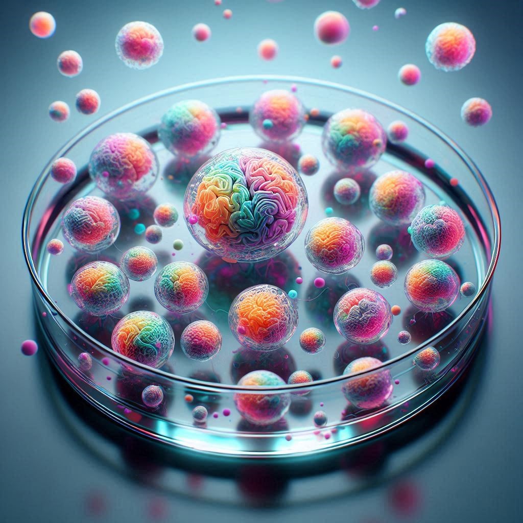
Cerebral organoids, often referred to as brain organoids or “minibrains,” are three-dimensional structures derived from human stem cells. These organoids mimic key aspects of brain development and function, providing a revolutionary tool for studying neurological diseases, brain development, and the effects of various drugs and treatments.
Formation and Development of Cerebral Organoids
The creation of cerebral organoids involves differentiating human pluripotent stem cells (hPSCs) into neural progenitor cells, which then self-organize into structures resembling the human brain. This process is supported by specific culture conditions and scaffolding proteins that facilitate the growth and organization of these cells into complex, multilayered tissues.
Cellular Composition and Structure
Cerebral organoids contain a diverse array of cell types found in the human brain, including neurons, glial cells, astrocytes, and microglia. These cells are organized into regions that reflect the anatomical and functional characteristics of the developing brain. For instance, brain organoids can form distinct layers resembling the cortical plate and exhibit region-specific neuronal activity.
Detailed Methodologies for Different Types of Cerebral Organoids
Cerebral organoids are a cutting-edge tool in neuroscience, offering insights into brain development and disease. Developing specific types of cerebral organoids, such as forebrain, midbrain, and hypothalamic organoids, requires tailored methodologies. Here, we provide detailed protocols for generating these brain-region-specific organoids, highlighting the necessary patterning factors and culture conditions.
1. Forebrain Organoids
Forebrain organoids mimic the development and structure of the human forebrain, a critical region responsible for cognitive functions and voluntary movement.
Materials and Methods:
- Stem Cell Culture: Start with human pluripotent stem cells (hPSCs).
- Embryoid Body Formation: Induce the formation of embryoid bodies (EBs) by culturing hPSCs in low-attachment plates.
- Neural Induction: After 4-5 days, transfer EBs to neural induction media containing dual SMAD inhibitors (e.g., Noggin and SB431542) to promote neural lineage commitment.
- Neural Patterning: To specify forebrain identity, supplement the media with Wnt inhibitors (e.g., IWR-1) and BMP inhibitors (e.g., LDN193189).
- Organoid Maturation: Embed the neural rosettes in Matrigel droplets and culture them in a spinning bioreactor with differentiation media containing BDNF, GDNF, and cAMP.
Diagram:
graph TD
    A[Human Pluripotent Stem Cells] --> B[Embryoid Bodies]
    B --> C[Neural Induction (Dual SMAD Inhibitors)]
    C --> D[Forebrain Patterning (Wnt and BMP Inhibitors)]
    D --> E[Organoid Maturation (Matrigel, Bioreactor)]
2. Midbrain Organoids
Midbrain organoids are essential for studying diseases such as Parkinson’s, as they contain dopaminergic neurons.
Materials and Methods:
- Stem Cell Culture: Begin with hPSCs.
- Embryoid Body Formation: Form EBs using low-attachment plates.
- Neural Induction: Induce neural differentiation with dual SMAD inhibitors.
- Midbrain Patterning: Add SHH (Sonic Hedgehog) and FGF8 (Fibroblast Growth Factor 8) to the media to promote midbrain identity.
- Organoid Maturation: Embed in Matrigel and culture in a spinning bioreactor. Supplement with ascorbic acid, GDNF, and BDNF to support dopaminergic neuron development.
Diagram:
graph TD
    A[Human Pluripotent Stem Cells] --> B[Embryoid Bodies]
    B --> C[Neural Induction (Dual SMAD Inhibitors)]
    C --> D[Midbrain Patterning (SHH and FGF8)]
    D --> E[Organoid Maturation (Matrigel, Bioreactor)]
3. Hypothalamic Organoids
Hypothalamic organoids are used to study metabolic disorders and neuroendocrine functions.
Materials and Methods:
- Stem Cell Culture: Culture hPSCs.
- Embryoid Body Formation: Generate EBs in low-attachment plates.
- Neural Induction: Use dual SMAD inhibitors to induce neural differentiation.
- Hypothalamic Patterning: Add SHH, FGF8, and BMP7 to the media to induce hypothalamic differentiation.
- Organoid Maturation: Culture the organoids in Matrigel droplets in a spinning bioreactor, with media supplemented with IGF-1 and T3 to promote hypothalamic neuron maturation.
Diagram:
graph TD
    A[Human Pluripotent Stem Cells] --> B[Embryoid Bodies]
    B --> C[Neural Induction (Dual SMAD Inhibitors)]
    C --> D[Hypothalamic Patterning (SHH, FGF8, BMP7)]
    D --> E[Organoid Maturation (Matrigel, Bioreactor)]
These detailed protocols for generating forebrain, midbrain, and hypothalamic cerebral organoids provide essential tools for advancing research in neurodevelopment and neurological disorders. By following these methodologies and using the appropriate patterning factors and culture conditions, researchers can create brain-region-specific organoids to study various aspects of brain function and disease.
Applications in Research
Studying Neurodevelopmental Disorders
Brain organoids have become invaluable in studying neurodevelopmental disorders such as autism spectrum disorder (ASD), microcephaly, and Rett syndrome. These models allow researchers to observe the developmental processes that may be disrupted in these conditions and to identify potential therapeutic targets.
Modeling Neurodegenerative Diseases
Cerebral organoids are also used to model neurodegenerative diseases like Alzheimer’s and Parkinson’s. By introducing disease-related mutations or environmental stressors, scientists can study the progression of these diseases and test the effects of new drugs in a controlled environment.
Drug Screening and Toxicology
One of the most promising applications of brain organoids is in drug screening and toxicology studies. These organoids provide a more accurate representation of human brain tissue compared to traditional two-dimensional cell cultures or animal models. This makes them ideal for testing the efficacy and safety of new pharmaceuticals.
Challenges in Vascularization and Solutions in Cerebral Organoids
Cerebral organoids have revolutionized neuroscience by providing a platform to study human brain development and diseases. However, a significant challenge in their development is the lack of vascularization, which limits their growth, survival, and functionality. This article explores the importance of vascularization in cerebral organoids, the current challenges, and the latest advancements in overcoming these obstacles.
Importance of Vascularization in Cerebral Organoids
Vascularization is crucial for the proper development and function of tissues, including cerebral organoids. Blood vessels supply oxygen and nutrients and remove metabolic waste, facilitating cell survival and function. In the context of cerebral organoids, vascularization is essential to:
- Enhance organoid survival: Providing a steady supply of oxygen and nutrients to prevent central necrosis.
- Improve functionality: Ensuring proper cell differentiation and functionality.
- Enable larger organoid size: Allowing the development of more complex and physiologically relevant structures.
Current Challenges in Vascularizing Cerebral Organoids
Despite the advancements in cerebral organoid research, vascularizing these structures poses several challenges:
- Lack of Endothelial Cells: Cerebral organoids typically lack endothelial cells, which are crucial for blood vessel formation.
- Limited Diffusion: Without blood vessels, nutrients and oxygen can only diffuse into the organoid from the culture medium, leading to central necrosis as the organoid size increases.
- Immature Vascular Networks: When endothelial cells are incorporated, the resulting vascular networks are often immature and do not fully mimic the complexity of native brain vasculature.
Advancements in Vascularization Techniques
Microfluidic Devices
Microfluidic devices have emerged as a promising tool for vascularizing cerebral organoids. These devices can simulate the dynamic environment of blood flow, promoting the formation of functional vascular networks within the organoids.
- Technique: Microfluidic chips with microchannels are used to culture organoids. These channels can be perfused with media containing growth factors that promote angiogenesis (the formation of new blood vessels).
- Benefits: Improved nutrient and oxygen delivery, enhanced cell survival, and more physiologically relevant vascular networks.
Co-Culture Techniques
Co-culturing cerebral organoids with endothelial cells or other supporting cell types has shown potential in promoting vascularization.
- Technique: Endothelial cells are mixed with neural progenitors during the initial stages of organoid formation or introduced into mature organoids. Growth factors and extracellular matrix components are used to support the co-culture environment.
- Benefits: Formation of blood vessel-like structures within the organoid, improved cell survival, and functionality.
Incorporation of Vascular Growth Factors
Adding vascular growth factors to the culture medium can stimulate the development of blood vessels within cerebral organoids.
- Technique: Supplementing the culture medium with growth factors such as VEGF (vascular endothelial growth factor), bFGF (basic fibroblast growth factor), and EGF (epidermal growth factor) to promote angiogenesis.
- Benefits: Enhanced vascular network formation, better nutrient and oxygen supply, and reduced central necrosis.
Successful Studies and Case Examples
Integration of Human Endothelial Cells
A study by Pham et al. (2018) demonstrated the successful incorporation of human endothelial cells into cerebral organoids. The researchers used a co-culture system with human induced pluripotent stem cell-derived endothelial cells, leading to the formation of vascular networks within the organoids. This approach significantly improved the survival and maturation of the organoids.
Microfluidic Vascularization
A research team led by Zhang et al. (2020) developed a microfluidic platform to vascularize cerebral organoids. The microfluidic device provided a continuous flow of culture medium, promoting the formation of mature blood vessels. The vascularized organoids exhibited improved growth, reduced necrosis, and enhanced neuronal differentiation.
In short, vascularization is a critical factor in the development and functionality of cerebral organoids. Overcoming the challenges of vascularization can lead to more physiologically relevant models for studying brain development and diseases. Techniques such as microfluidic devices, co-cultural methods, and the incorporation of vascular growth factors offer promising solutions. Continued advancements in these areas will enhance the potential of cerebral organoids in research and therapeutic applications.
Comparative Analysis of Cerebral Organoids and Traditional Models
Cerebral organoid research has rapidly advanced, offering new insights into human brain development and disease. However, traditional in vitro and in vivo models continue to play a crucial role in neuroscience. This article provides a comprehensive comparative analysis of cerebral organoids versus traditional models like 2D cell cultures and animal models, evaluating their strengths, limitations, cellular diversity, functional relevance, and scalability. We will also present case studies highlighting unique insights gained from cerebral organoids that traditional models could not provide.
Strengths and Limitations
Cerebral Organoids
Strengths:
- Human Relevance: Cerebral organoids are derived from human pluripotent stem cells, offering a more accurate model of human brain development and disease than animal models.
- 3D Structure: Unlike 2D cell cultures, cerebral organoids possess a three-dimensional structure, mimicking the complexity of the human brain’s architecture.
- Cellular Diversity: Organoids can develop various brain cell types, including neurons, astrocytes, and oligodendrocytes, reflecting the cellular diversity of the human brain.
- Genetic Manipulation: Organoids can be genetically modified to study the effects of specific genes on brain development and function.
Limitations:
- Lack of Vascularization: Cerebral organoids often lack blood vessels, limiting their growth and functional maturation.
- Immature State: Organoids typically represent early developmental stages and may not fully mature to resemble adult brain tissue.
- Variability: There can be significant variability between organoids, making reproducibility a challenge.
Traditional Models
2D Cell Cultures:
- Strengths:
- Simplicity: Easy to culture and manipulate, making them ideal for high-throughput screening.
- Cost-Effective: Less expensive and resource-intensive compared to 3D models and animal studies.
- Limitations:
- Lack of 3D Architecture: Do not mimic the complex 3D structure of the brain.
- Limited Cellular Diversity: Typically consist of a single cell type, lacking the cellular diversity of brain tissue.
Animal Models:
- Strengths:
- Complex Systems: Provide a whole-organism context, allowing the study of systemic interactions.
- Functional Studies: Enable behavioral and physiological studies that are not possible in vitro.
- Limitations:
- Species Differences: Differences between animal and human brains can limit the translational relevance of findings.
- Ethical Concerns: Animal studies raise ethical issues and are subject to stringent regulatory requirements.
Functional Relevance and Scalability
Functional Relevance
Cerebral Organoids:
- Organoids can recapitulate key aspects of brain development and function, such as neuronal differentiation, synapse formation, and network activity. However, their lack of vascularization and limited maturation can impact their functional relevance.
2D Cell Cultures:
- While useful for studying basic cellular processes, 2D cultures fail to replicate the complex interactions and 3D architecture of brain tissue, limiting their functional relevance.
Animal Models:
- Provide valuable insights into brain function and disease mechanisms, allowing for behavioral studies and complex physiological assessments. However, species differences can limit the relevance of these findings to humans.
Scalability
Cerebral Organoids:
- Scalable to an extent but require specialized equipment and expertise. High variability and limited reproducibility remain challenges.
2D Cell Cultures:
- Highly scalable and amenable to high-throughput screening, making them ideal for drug discovery and basic research.
Animal Models:
- Less scalable due to ethical concerns, regulatory requirements, and higher costs. Suitable for detailed, hypothesis-driven studies but not for large-scale screening.
Case Studies
Autism Spectrum Disorder (ASD)
Cerebral Organoids:
- Researchers used cerebral organoids derived from patients with ASD to identify disrupted neurodevelopmental processes. These organoids exhibited abnormal neuronal migration and altered synaptic function, providing insights that traditional models failed to capture.
Zika Virus Infection
Cerebral Organoids:
- Cerebral organoids were instrumental in studying the impact of Zika virus on brain development. The organoids revealed that the virus targets neural progenitor cells, leading to microcephaly, a finding that was challenging to reproduce in animal models due to species differences.
In Short Cerebral, organoids offer a unique and valuable model for studying human brain development and disease, complementing traditional in vitro and in vivo models. While they provide significant advantages in terms of human relevance and cellular diversity, challenges such as lack of vascularization and variability remain. Traditional models, including 2D cell cultures and animal models, continue to play a critical role, offering simplicity, scalability, and the ability to study complex systemic interactions. By leveraging the strengths of each model and addressing their limitations, researchers can gain a more comprehensive understanding of the brain and its disorders.
Applications in Neurodevelopmental Disorders
Cerebral organoids have emerged as powerful tools for studying neurodevelopmental disorders, providing insights that traditional models often cannot. This article delves into specific examples of how cerebral organoids are used in researching neurodevelopmental disorders such as autism spectrum disorder (ASD) and schizophrenia. It includes details on the genetic modifications used, observed phenotypes, and how these findings are advancing our understanding and potential treatments.
Autism Spectrum Disorder (ASD)
Autism Spectrum Disorder is a complex neurodevelopmental condition characterized by social, communication, and behavioral challenges. The use of cerebral organoids has provided valuable insights into the underlying mechanisms of ASD.
Case Study: Genetic Modifications and Observed Phenotypes
Genetic Modifications:
- Researchers have used induced pluripotent stem cells (iPSCs) derived from patients with ASD to create cerebral organoids. Common genetic modifications include mutations in genes such as CHD8, SHANK3, and PTEN, which are associated with ASD.
Observed Phenotypes:
- CHD8 Mutations: Organoids with CHD8 mutations exhibit enlarged size, reflecting macrocephaly observed in some ASD patients. They also show altered neuronal differentiation and increased proliferation of neural progenitor cells.
- SHANK3 Mutations: Organoids with SHANK3 mutations demonstrate synaptic dysfunction, including reduced synapse formation and impaired neuronal connectivity. This mirrors the synaptic deficits often seen in ASD.
- PTEN Mutations: Organoids with PTEN mutations display abnormal cortical development, including disrupted layering and altered cell migration. These phenotypes are consistent with the brain abnormalities found in some individuals with ASD.
Advances in Understanding and Treatment
Using cerebral organoids to model ASD has led to several advancements:
- Mechanistic Insights: Researchers have identified key pathways and cellular processes disrupted in ASD, such as altered Wnt signaling and increased oxidative stress.
- Drug Screening: Organoids provide a platform for testing potential therapeutic compounds. For instance, IGF-1 (insulin-like growth factor 1) has shown promise in rescuing some of the cellular abnormalities in ASD organoids.
Schizophrenia
Schizophrenia is a severe neuropsychiatric disorder characterized by hallucinations, delusions, and cognitive impairments. Cerebral organoids offer a unique model to study the early developmental changes associated with schizophrenia.
Case Study: Genetic Modifications and Observed Phenotypes
Genetic Modifications:
- Researchers have used iPSCs from patients with schizophrenia to generate cerebral organoids. Key genetic modifications include mutations in genes like DISC1, NRG1, and CACNA1C, which are implicated in the disorder.
Observed Phenotypes:
- DISC1 Mutations: Organoids with DISC1 mutations exhibit abnormal neural progenitor cell proliferation and migration, leading to disrupted cortical layering. These findings are consistent with the cortical abnormalities observed in schizophrenia patients.
- NRG1 Mutations: Organoids with NRG1 mutations show altered synaptic function and reduced dendritic spine density, reflecting synaptic deficits seen in schizophrenia.
- CACNA1C Mutations: Organoids with CACNA1C mutations display impaired neuronal connectivity and altered calcium signaling, which are linked to the cognitive and functional impairments in schizophrenia.
Advances in Understanding and Treatment
Cerebral organoids have advanced our understanding of schizophrenia in several ways:
- Pathophysiological Mechanisms: Studies using organoids have identified disrupted pathways, such as altered GABAergic signaling and impaired mitochondrial function, contributing to schizophrenia.
- Therapeutic Targets: Organoids facilitate the identification of novel therapeutic targets. For example, treatments aimed at restoring calcium signaling in CACNA1C-mutant organoids have shown potential benefits.
In short, the use of cerebral organoids in studying neurodevelopmental disorders like autism and schizophrenia has provided significant insights into the molecular and cellular mechanisms underlying these conditions. By modeling patient-specific genetic mutations and observing the resulting phenotypes, researchers can better understand the pathophysiology of these disorders and identify potential therapeutic targets. Continued advancements in cerebral organoid technology will likely lead to further breakthroughs in the diagnosis and treatment of neurodevelopmental disorders.
Technological Advances and Enhancements
Recent advancements have enabled the creation of more complex brain organoids that can simulate interactions between different brain regions. For example, researchers form assembloids by fusing organoids from various brain regions to study the crosstalk between different neuronal populations. Additionally, scientists are making efforts to incorporate blood vessels and immune cells into organoids to better mimic the in vivo environment.
Ethical Considerations
While cerebral organoids offer numerous benefits for research, they also raise ethical concerns. As these organoids become more complex, questions about their potential for sentience and the ethical implications of their use in research have been raised. It is crucial for the scientific community to address these concerns and establish guidelines for the ethical use of brain organoids.
Future Directions
The future of cerebral organoids is promising, with ongoing research focused on improving their complexity and functionality. Scientists aim to create more advanced models that can replicate human brain development and disease more accurately. These advancements could lead to new therapies for neurological disorders and reduce the reliance on animal models in research.
Culmination
Cerebral organoids represent a significant leap forward in neuroscience research. Their ability to model human brain development and disease in a controlled environment opens new avenues for understanding the brain and developing treatments for neurological disorders. As technology advances, the potential applications of brain organoids will continue to expand, offering new hope for patients and researchers alike.
Recommended Reading
- Stem Cell Research & Therapy – A beginner’s guide on the use of brain organoids for neuroscientists
- Live Science – Cerebral organoids: What are lab-grown ‘minibrains’?
- Springer – Advances and Applications of Brain Organoids
This comprehensive overview provides insights into the fascinating world of cerebral organoids, highlighting their formation, applications, and future potential in advancing neurological research.